Image -
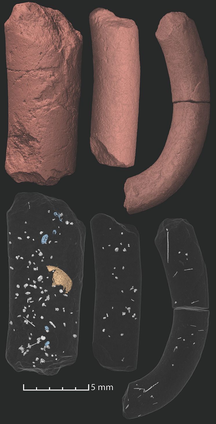
Fossil droppings
The fossil droppings were scanned using synchrotron microtomography. This works in a similar way to a CT-scanner in a hospital but with much stronger x-ray beams, making it possible to image the contents of fossils in three dimensions. The scans revealed many microscopic food remains including foraminifera (small amoeboid protists with external shells), small shells of marine invertebrates and possible remains of polychaete worms.
Qvarnström et al.
- License:
- Media Use
The content may be downloaded by journalists, bloggers, columnists, creators of public opinion, etc. It can be used and shared in different media channels to convey, narrate, and comment on your press releases, posts, or information, provided that the content is unmodified. The author or creator shall be attributed to the extent and in the manner required by good practice (this means, for example, that photographers should be attributed).
- By:
- Qvarnström et al.
- File format:
- .jpg
- Size:
- 2011 x 3949, 3.25 MB